 |
 |
||
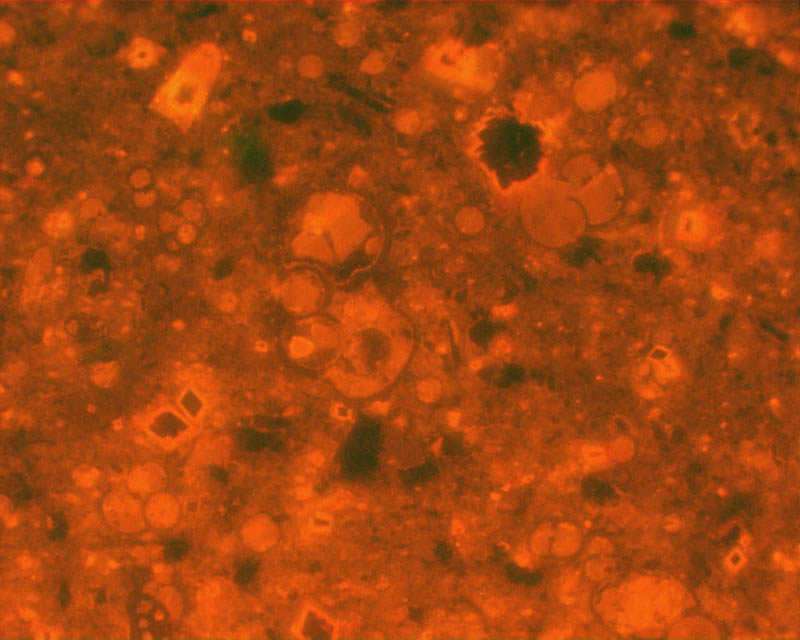
Backscatter image of sample yax315. The scale from left to right is 5mm. Dark grey areas are dolomite (Mg, CaCO3), bright is calcite (CaCO3) and dark is empty porespace or silicate. The region in pink is enlarged on the right (top) |
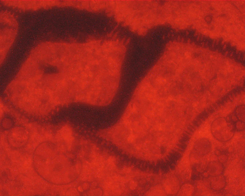
Enlargement of colored area of image on the left. The bright areas are pore-filling blocky calcite (the filling is one single crystal). The calcite encloses authigenic low-temperature quartz idomorphic crystals, as is show on the enlargement here below, where the different chemical elements are X-ray mapped on the electron microprobe. Below enlargement of coloured area | ||
 |
|||
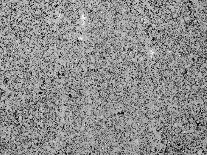
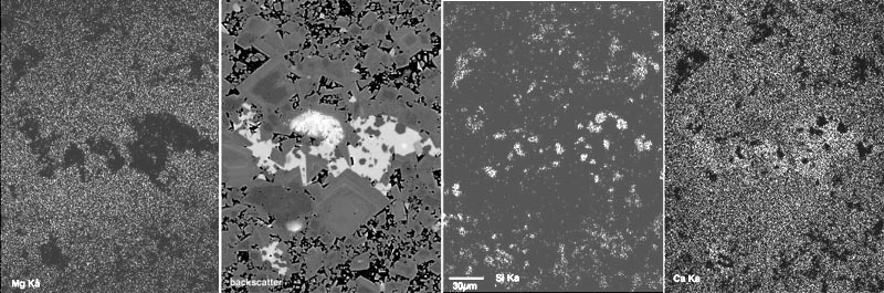
X-ray map of the Mg Ka peak, showing clearly that dolomite is the dominant composition. The large dark area does not show any Mg Ka peak, because the region is pure CaCO3, normal for low-temperature calcite |
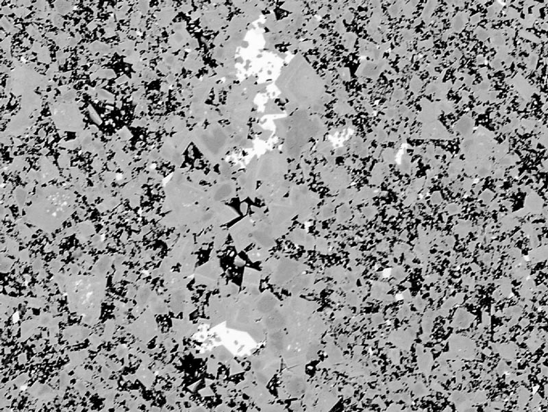
Backscatter image of the X-ray mapped region. Clearly visible is that all dolomite crystals have the same complex zoned idiomorphic overgrowth, the porespace is dark because it is either empty, or filled with clayminerals | X-ray map of the Si Ka peak, showing the quartz crystals, and the clay mineral filling of the porespaces | X-ray map of the Ca Ka peak. Calcite CaCO3 shows stronger reflection than dolomite (Mg, CaCO3). Quartz and the porespaces do not show Ca reflections. This rock is not a micrite |
 |
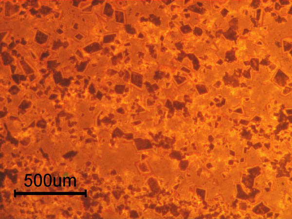
Sample Yax315, in the interval qualified as "laminated micritic limestone" by(op.cit. Keller and Stinnesbeck). Overlay images of Cathode luminescence and transmitted light |
| Next - - Previous | |
|
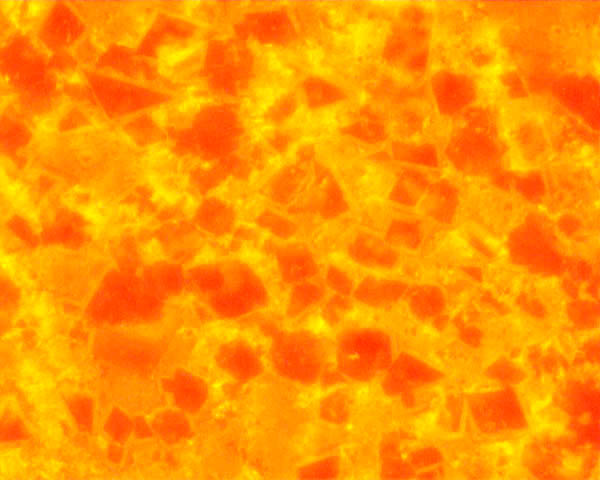
Sample Yax315, in the interval qualified as "laminated micritic limestone" by(op.cit. Keller and Stinnesbeck). Overlay images of Catode luminescence and backscatter SEM image |
| Next- - Previous | |
 |
Sample Yax315, in the interval qualified as "laminated micritic limestone" by(op.cit. Keller and Stinnesbeck). Overlay images of Transmitted light and backscatter SEM image |
| Next - - Previous |
 |
Sample Yax310, When you move your mouse back and forth over the image you will see a quick comparison of Backscatter electron image and transmitted light image. Same area as the image above |
| Next - - Previous |
 |
Sample Yax310. When you move your mouse back and forth
over the image you will see a quick comparison of cathode luminescence and
Backscatter electron image. Same area as the image above |
|
|
|
Sample Yax310, When you move your mouse back and forth over the image you will
see a quick comparison of cathode luminescence and transmitted light images
of exactly the same area at the same magnification. |
|
|
 |
Sample Yax309, change over from Cathode Luminescence to transmitted light |
|
 |
|||||
| Similar TL image published by Keller et al (2004, 2004a) | ||||||
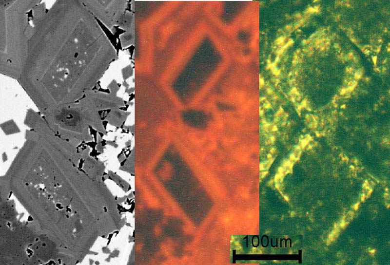
| (A) from Keller et al (PNAS and MAPS (here) | (B) Tl image of dolomite crystals, in an arrangement that greatly resembles A, so-called" foraminiferal chambers" | (C) CL image of same area as B, bright luminescence is due to high Mn content | (D) backscatter SEM image of same area as B and C. The dolomite core is porous and filled with (white) calcite.Also the porespace between the dolomite crystals is filled with sparitic calcite | |||
| All images of the same dolomite crystals in sample yax310. (B=Tl , C=CL and D= SEM) (see detailed comparison) top | ||||||
 |
 |
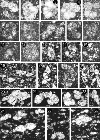
Images published by Keller et al (2004, left 2004b, right) 1-12 are legitimate foraminifers (see here) because they have nice curved
teswalls, with apertures and a good testwall structure on Cl and SEM images. Click on the pink area to compare these shapes in other imaging methods Keller, G., Adatte, T., Stinnesbeck, W., Rebolledo-Vieyra, M., Urrutia Fucugauchi, J., Kramar, U., and Stuben, D., 2004, Chicxulub impact predates the K-T boundary mass extinction: PNAS, v. 101, no. 11, p. 3753-3758. |
|
Images published by (Keller 2004, 2004a) |
||
| SEM, CL and TL images of two dolomite crystals | last updated: July 10, 2018 |
||
| Micritic limestone with foraminifers? |
|||
| Foraminifers from the Scaglia Rossa | 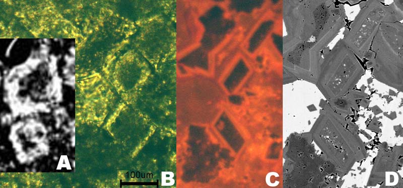 |
||
| Foraminifers and dolomite crystals from ODP leg 1049 | |||
| Cl and Tl compared in sample 315 | rollover images of Tl and Cl of sample yax309 |
| Move your mouse across image to see comparison between two imaging methods |
Forum on the Geological Society debate, forams and dolomite crystals |