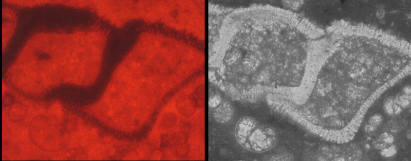
Comparison of Transmitted light (TL) and Cathodeluminescense (CL) microscopy.
Upper Maastrichtian of Scaglia Rossa, Piobbico, compared to sample 20
of Keller et al.
|
Comparison of Transmitted light (TL) and Cathodeluminescense (CL) microscopy. |
last updated: November 10, 2004 | |
 |
|
| CL (cf. Globotruncana stuarti) dark wall, light interior | TL, same foraminifer, light wall, dark interior |
| The reason being that the foraminiferal rim is a pure calcite which almost not reflective (=dark in CL), and the chambers are filled with 'dirty' micrite, which is CL reflective (=light) due to higher Fe content. This is also in other micrite settings the case, (see example from ODP site 1049, Blake Nose). | |
 |
 |
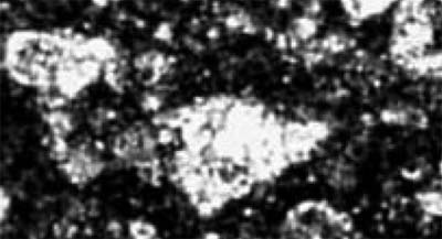
| Images from Yaxcopoil-1 believed by Keller to be foraminifers. Although the outline may be somewhat reminiscent of the foraminifer shown above, there are no details visible to positively identify them. Note that the outline of each of the "chambers", can be easily created by a cross-section through a dolomite crystal, that are abundantly present in these so-called "micritic limestone" intervals (see these images). The solution to this problem is straightforward: These shapes should be subjected to SEM backscatter (+X-ray mapping) and Cathodeluminescense (CL) image analysis, and then the difference between dolomite crystals and a calcite-filled foraminiferal shell will show up clearly. | |